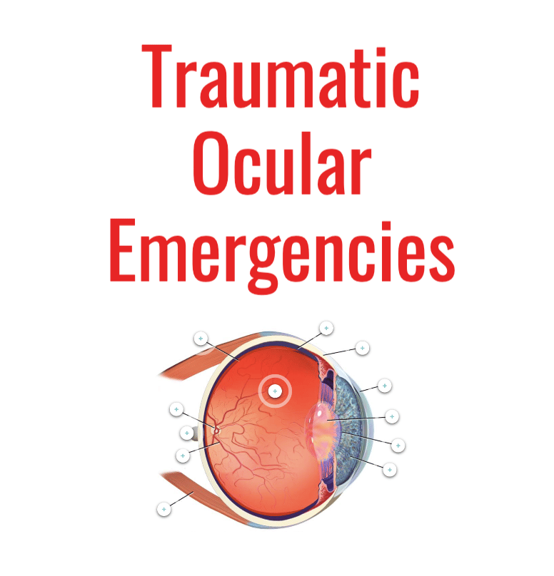
The eye and its surrounding structures are highly complex, and an injury to this important body part can lead to permanent vision loss and disability. Because of these implications, many nurses are hesitant to care for patients with ocular injuries.
While traumatic eye injuries can be distracting, they require rapid assessment to ensure that both blood supply and optic nerve function are maintained. Following are eight ocular injuries that trauma and emergency nurses should understand and be prepared to encounter.
1. Corneal abrasion
As the name implies, corneal abrasions are localized to the cornea. These abrasions on the surface of the eye can be visualized under fluoroscopy using fluorescein dye and a Wood’s lamp (or another blue light source). The dye can be administered as eye drops or using fluorescein strips that are gently placed on the eye itself.
Treatment for corneal abrasions may include ophthalmic ointment, antibiotics and/or anesthetic drops.
2. Chemical splash
Patients that experience a chemical splash to the eye will most likely require copious irrigation. This can prove to be a challenge, especially for pediatric patients.
Eyes may be irrigated using a commercial eyewash station, IV tubing held over the eye, a rigid silicone contact lens or a large-volume catheter-tip syringe. (Refer to your hospital’s policies and procedures for eye irrigation.)
Many providers use an irrigation solution with a pH of 7.0 to match the normal pH of the eye.
3. Foreign body in eye
If a foreign body cannot be flushed out via irrigation, it should be stabilized as much as possible to prevent further damage to the eye. Stabilization measures may include:
- Covering the eye with a dressing that prevents further contamination but does not touch the eye itself.
- Covering both eyes if there is a chance the injury will worsen based on the location of the object and the patient’s congruent eye movement.
- Minimizing environmental stimulation and encouraging the patient to maintain a forward-facing gaze.
- Instilling eye drops and/or eye ointments as directed.
4. Open globe injury
Any full-thickness injury to the sclera (the white part of the eye), the cornea or both is considered an open globe injury. This can lead to vision loss and create an opening for bacterial contaminants. It can also lead to a complete globe rupture in which the eye loses its shape.
Treatment for an open globe injury may include:
- Tetanus toxoid administration
- Radiology imaging of surrounding structures
- Laboratory tests in preparation for surgery
- Antibiotic administration
5. Orbital fracture
The orbital socket contains several fused bones that protect and support the eye. Nurses should suspect orbital fracture in any patient who has experienced direct eye trauma or anyone presenting with a large penetrating object.
- Orbital rim fracture is a break in any portion of the bones of the rim of the eye. These bones require significant force to fracture. This fracture should lead to suspicion of optic nerve injury.
- Orbital floor fracture results when compression and break of the orbital rim results in a break in the orbital floor as well.
- Blowout fracture is a break in the walls or floor of the orbit. This fracture can lead to ocular entrapment and significant visual difficulties.
6. LeFort fracture
LeFort fractures result from direct trauma to the face, often from motor vehicle collisions. There are three types of LeFort fracture and they have different implications for eye injury:
- LeFort I fractures are isolated to the upper maxillary region above the upper teeth. This fracture does not impact the eyes but does form a floating palate.
- LeFort II fractures indirectly impact the eyes, involving the nose and inferior orbital rim. Patients with these fractures have a floating maxilla.
- LeFort III fractures involve a complete craniofacial separation. These injuries prevent the eyes from being adequately supported.
7. Retrobulbar hematoma
A retrobulbar hematoma is a hematoma that forms from a hemorrhage deep within the posterior orbital area. Retrobulbar hematoma is rare, but it is an emergent condition that requires rapid treatment. Expanding retrobulbar hematomas can cause significant intraocular compartment pressure and loss of vision.
Symptoms of retrobulbar hematoma may include:
- Severe pain in the affected eye
- Periorbital swelling and bruising
- Exophthalmos (bulging out of the eye)
- Loss of vision in the affected eye
- Progressive loss of ocular movement
8. Maxillofacial trauma with object impalement
Patients involved in significant traumatic events (e.g., high-speed crashes, assaults and/or fall injuries) have the potential for maxillofacial trauma with object impalement. Object impalements can be very distracting for the entire trauma team, and the necessary care is complex.
- Patients with significant facial trauma require continuous reassessment of their airway, breathing status, circulation and neurovascular status.
- Any large, impaled object should be stabilized until surgical removal.
- Penetrating objects that impact the eye and surrounding tissues carry a high risk of infection. Apart from the usual ABC prioritization, nurses should be prepared to decrease the risk for any additional external contamination. A washout and debridement may be necessary on more than one occasion while the patient is admitted.
- The patient should also be assessed for signs of a CSF leak.
Patients arriving at a facility that does not perform complex maxillofacial surgeries should be rapidly transferred to a higher level of care as soon as the injury is recognized. According to the 2022 edition of Resources for Optimal Care of the Injured Patient, both adult and pediatric Level I trauma centers must have the capability to diagnose and manage acute facial fractures of the entire craniomaxillofacial skeleton.


